Showing posts with label methods and techniques. Show all posts

Autoradiography

Autoradiography
Autoradiography
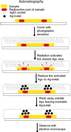
Autoradiography produces an image formed by a substance’s own radioactivity when exposed to a photographic film. This technique is often used for investigation of biological processes.

In 1896 Antoine-Henri Becquerel was working with rocks containing uranium ore. By chance, he put one rock sample into a dark drawer on top of a box of unexposed photographic film. When the film later was developed, it showed a clear outline of the uranium rock.

Evidently, some radiation had been emitted from the rock, penetrated through the wrapping paper, and exposed the film inside. An autoradiograph, that is, an image produced by radioactivity, was visible on the film. Autoradiography, much refined, is now a valuable technique for investigating biological processes.

Hungarian chemist Georg von Hevesy pioneered the use of radioactive tracers in biological research in the 1920’s, and two developments in the 1930’s greatly expanded their use. Firstwas the discovery of induced radiation by Frédéric Joliot-Curie and Irène Joliot-Curie, which raised the exciting possibility that artificially created radioactivity could be induced in almost any element found in nature.

Chromatography

Chromatography
Chromatography
Chromatography is a method of separating the components of a mixture over time. Chromatography has allowed for the discovery of many specialized pigments, including at least five forms of chlorophyll.

Chromatography was first described in 1850 by a German chemist, Friedlieb Ferdinand Runge. It was not until the early twentieth century, however, that Mikhail Semenovich Tsvet became the first to explain the phenomenon and methods of this analytical tool.

Chromatography and Photosynthesis

Tsvet’s chromatography of plant leaf pigments prompted scientific investigations of photosynthesis—the all-important biochemical reaction that transforms inorganic to organic energy and therefore is at the base of most life. Chromatography has revealed that many different pigments, not only green ones, are simultaneously present in leaves.

Cladistics

Cladistics
Cladistics
Cladistics is a quantitative method of classification of plants that attempts to recover evolutionary relationships, based on observable characters.

Since the dawn of history, humans have classified plants. In primitive cultures classifications were by economic use, such as food, clothing, medicine, and shelter. Later the form (morphology) of a plant became important, for example, trees, shrubs, or herbs.

Carolus Linneaus considered the similarity of floral parts to be critical, and this formed the basis of his classification system. Each of these systems is said to be “artificial.” That is, the classification was solely for a human purpose and did not attempt to indicate genetic relationships between plants.

Dendrochronology

Dendrochronology
Dendrochronology

Dendrochronology is the science of examining and comparing growth rings in both living and aged woods to draw inferences about past events and environmental conditions.

In forested regions with seasonal climates, trees produce a growth ring to correspond with each growing season. At the beginning of the growing season, when conditions are optimum, the vascular cambium produces many files of large xylem cells that form wood.

As the conditions become less optimal, the size and number of cells produced decreases until growth stops at the end of the growing season. These seasonal differences in size and number of cells produced are usually visible to the unaided eye.

Electrophoresis

Gel electrophoresis
Gel electrophoresis

Electrophoresis is a biochemical technique used to separate charged molecules in an electric field. Gel electrophoresis is one of the most common forms of this method, used to separate DNA, proteins, enzymes, and other molecules from the cell for laboratory investigation and manipulation.

Electrophoresis is widely used to separate, visualize, or purify charged biological molecules such as deoxyribonucleic and ribonucleic acids (DNA and RNA) and proteins, including enzymes.

It is also used to estimate the size of DNA fragments and the molecular weight of proteins. Most biological molecules are electrically charged in solution; hence, when subjected to an electric field, they migrate as zones toward an electrode (a terminal source of electricity) of opposite electrical polarity.

Fluorescent Staining of Cytoskeletal Elements

Fluorescent Staining of Cytoskeletal Elements

Fluorescent antibody staining is a precise technique for marking elements of the cytoskeleton so they become visible under a microscope. The technique has revealed much about the location and functions of the cytoskeleton within a cell. It also offers some of the most visually appealing of the microscopic images available to biologists.

Cells require an internal system of fibers in order to maintain and change their shape. Perhaps the most straightforward function of the cytoskeleton is to provide cell support, a scaffolding that gives each cell a distinctive three-dimensional shape.

Without such support every cell would be shaped like a fried egg. The fibers found in the cytoskeleton serve other functions as well: for example, as rails along which substances shuttle from one part of the cell to another.