Nucleic acids are the genetic material of cells, including DNA and the various types of RNA.
Nucleic acids were discovered in the mid-nineteenth century, but their role as genetic material was not substantiated until the mid twentieth century. When chromosomes were discovered at the beginning of the twentieth century, they were quickly identified as the genetic material of the cell. Chromosomes were found to be composed of nucleic acids and proteins.
Through the experiments of Fred Griffith on transformation in pneumonia bacteria and the work of Alfred Hershey and Martha Chase on bacteriophages, by 1952 most biologists recognized deoxyribonucleic acid (DNA) as containing the genes. James Watson and Francis Crick provided the capstone to science’s initial understanding of nucleic acids when they determined the double helix structure of DNA in 1953.
  |
Heredity is the process by which the physical traits of an organism are passed on to its offspring. At the molecular level, DNA contains the information necessary for the transmission of genetic characteristics from one generation to the next, as well as the information required for the new organisms to growand to live. DNA is the chemical basis of heredity and provides the synthesis of new proteins, such as enzymes.
DNA and RNA
There are two types of nucleic acids within cells, the single-stranded ribonucleic acid (RNA) and the double-stranded DNA. Each kind has specific roles. DNA was isolated in 1869 by German chemist Friedrich Miescher.
The substance that Miescher found was white, sugary, and slightly acidic, and it contained phosphorus. Because it occurred only within the nuclei of cells, he called it “nuclein.” The name was later changed to deoxyribonucleic acid, to distinguish it from ribonucleic acid, which is also found in cells.
In eukaryotic cells, DNA is present in the chromosomes of the nucleus and within the mitochondria and chloroplasts. Bacteria, yeasts, and molds, in addition to the chromosomes, contain circular strands of DNA, called plasmids, within the cytoplasm of their cells.
Plasmids are relatively small, circular strands of DNA that exist independently of the chromosome. Plasmids typically have only twenty-five or thirty genes, which are not essential to the host cell but often confer antibiotic resistance, the ability to pass DNA to other bacterial cells, and other useful functions.
Some plasmids are only found as single copies, whereas others occur as many copies. The mitochondria and chloroplasts of eukaryotic cells are self-replicating and contain a tiny circular chromosome (DNA) resembling a plasmid of a bacterium.
Viruses (minute parasites that infect specific hosts) contain only one type of nucleic acid—either DNA or RNA, never both, and the DNA or RNA can be single- or double-stranded. The DNA of some viruses can integrate into the DNA of the host cell. In this state, the viral DNA replicates as the host DNA replicates.
The genetic apparatus of a virus, whether RNA or DNA, is much the same as that of bacteria but is far less complex. Even large viruses (such as the pox virus) have only a few hundred genes. Smaller viruses (such as the polio virus) have considerably fewer.
Chemical Structure
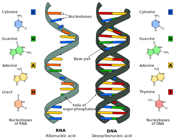
Both DNA and RNA are long-chained polymers made up of nucleotides. The nucleotides, in turn, are made up of a nitrogenous base, a ribose or deoxyribose sugar, and a phosphate. All the bases of DNA and RNA are heterocyclic amines.
Two, adenine and guanine, are called purines; the other three, cytosine, thymine, and uracil, are called pyrimidines. The two purines and one of the pyrimidines, cytosine, occur in both RNA and DNA. Uracil is found only in RNA, while thymine occurs only in DNA.
The purines are nine-membered heterocyclic rings with nitrogen occurring in place of carbon at several positions. Adenine and guanine differ in the functional groups attached to them.
The pyrimidines are six-membered heterocyclic rings,with nitrogen in place of two of the carbon atoms. Like the purines, the three pyrimidines also differ in the specific functional groups attached to them.
The ribose sugars are made up of a five-membered heterocyclic ring containing one oxygen atom between carbons one and four. A fifth carbon (number five) is not part of the ring and is bonded to carbon number four.
Along with hydrogen atoms attached to each carbon, there is one hydroxyl group (OH) attached to each of the four heterocyclic carbon atoms in the ribose sugar of RNA. (The fifth carbon has a phosphate group attached to it.) The sugar of DNA is called D-deoxyribose because a hydroxyl group is missing from the second carbon, having been replaced by a hydrogen atom—thus the name deoxyribonucleic acid.
The sugar-base combination is called a nucleoside. The purines are linked to carbon one of the sugars with the nitrogen at position one. The nucleoside of guanine and ribose is guanosine; it is adenosine for adenine and D-ribose.
The pyrimidines of RNA, when attached to ribose, are uridine and cytosine. In DNA the nucleoside names are deoxyadenine, deoxyguanosine, deoxythymidine, and deoxycytidine.
Nucleotides are phosphate esters of nucleosides. In these molecules, a phosphate group (phosphoric acid) is attached to carbon five (called the 5′ carbon) of the sugar (ribose or deoxyribose) in the nucleoside.
Nucleotides are named by combining the name of their nucleoside with a word describing the numbers of phosphates attached to it. Guanosinemonophosphate, for example, is the name of the phosphate ester of guanosine, which is often abbreviated as GMP.
Individual nucleotides also occur in cells. These free nucleotides usually exist as diphosphates or triphosphates. Examples of these are adenosine diphosphate (ADP) and adenosine triphosphate (ATP). ATP is the universal energy source for the anabolic processes of all cells, including the formation of the DNA and RNA.
DNA Structure and Function
DNA can be an extremely long molecule that is tightly wound within the nuclei of eukaryotic cells and within the cytoplasm of prokaryotic cells. Nuclear DNA is linear, whereas prokaryotic DNA is circular. (If the DNA in a human cell could be stretched out, it would measure roughly 2 meters, or 6 feet long; bacterial DNA would be about 1.5 millimeters long, or just over 0.5 inch.)
Using a typical lily as a point of reference, DNA is packaged into twenty-four chromosomes, twelve of which are contributed by the pollen and twelve by the egg. Every cell derived from the fertilized egg (zygote) will have exactly the same amount of DNA containing exactly the same genetic information.
Within the cytoplasm, several mitochondria (the sites of respiration) and chloroplasts (the sites of photosynthesis) are found, both of which contain their own DNA, which is circular and resembles prokaryotic DNA in many respects.
DNA is a double-stranded spiral; its shape is called the double helix. Structurally, it may be compared to a ladder, with the rails or sides of the ladder consisting of alternating deoxyribose sugar and phosphate molecules connected by phosphodiester bonds between the 5′ carbon of one sugar and the 3′ carbon of the other.
The rungs of the ladder consist of purine (adenine and guanine, often abbreviated as Aand G, respectively) and pyrimidine (cytosine and thymine, often abbreviated as C and T, respectively) building blocks from the opposite strands, held together by hydrogen bonds.
The building blocks pair with each other consistently in what are called complementary pairs: Adenine always pairs with thymine with two hydrogen bonds, and cytosine always pairs with guanine with three hydrogen bonds. Consequently, the attraction between cytosine and guanine is stronger than that between adenine and thymine.
Because of this arrangement, the sequence of the purine and pyrimidine building blocks on one strand is complemented by the sequence of building blocks on the other strand.
The specificity of the base pairing between the two strands allows strands to fit neatly together only when such pairing exists. Each DNA strand has a 5′ end with a hydroxyl group attached to the 3′ carbon of a deoxyribose sugar.
When connected, the two strands are actually in an opposite orientation and are referred to as being anti parallel. This is best observed by looking at one end of the double-stranded molecule. One strand terminates with a 5′ phosphate group and the other with a 3′ hydroxyl group.
The specific nucleotide composition in a species is essentially constant but can vary considerably among organisms. Regardless, the amounts of adenine and thymine are always the same, as are the amounts of guanine and cytosine because of the required complementary pairing.
Due to the greater strength of G-C bonds, organisms with a high GC content have DNA that must be heated to a higher temperature to denature, or separate, the strands. Some bacteria that live in hot springs have an especially high GC content.
The instructions contained within the DNA molecules occur in segments called genes. Most genes instruct the cell about what kind of polypeptide (molecule composed of amino acids used to make functional proteins) to manufacture.
These polypeptides lead to the formation of enzymes and other proteins necessary for survival of the cell. Other genes are important in coding for the production of antibodies, RNA, and hormones.
RNA Types
The DNA acts as a template to make three kinds of RNA: messenger RNA (mRNA), transfer RNA (tRNA), and ribosomal RNA (rRNA). Each kind of RNA has a specific function. RNA is not found in chromosomes and is located elsewhere in the nucleus and in the cytoplasm.
The largest and most abundant RNA is rRNA. Between 60 and 80 percent of the total RNA in cells is rRNA, and it has a molecular weight of several million atomic mass units. The rRNA combines with proteins to form ribosomes, which are the sites for the synthesis of new protein molecules. About 60 percent of the ribosome is rRNA, and the rest is protein.
Although single-stranded, rRNA molecules fold into specific functional shapes that involve the pairing of portions of the molecule to form double stranded regions. The precise shape of rRNAs is important for their function, and some of them actually have catalytic properties, just as enzymes do. RNAs of this type are sometimes calledribozymes.
Molecules of mRNA carry the genetic information from DNA to the ribosomes. The process of converting the DNA code of a gene into an mRNA is called transcription. When attached to the ribosomes, mRNAs direct protein synthesis in a process called translation.
The size of the mRNA molecule depends upon the size of the protein molecule to be made. In prokaryotes (such as bacterial cells), as well as in mitochondria and chloroplasts, mRNAs are ready to take part in translation even while transcription of the remainder of the mRNA is taking place.
In eukaryotes (cells of most other forms of life), them RNAs transcribed from nuclear DNA are initially much larger than they are later, when they participate in translation. These mRNAs must be processed to removed large pieces of noncoding RNA, called introns, and to modify both ends of the mRNA in specificways.
After introns are removed, the remaining codon regions, called exons, are spliced together by splicesomes (a complex system composed of proteins and small RNAs). Once all the modifications are complete, them RNA is ready to be translated. A small number of mRNAs are translated in the nucleus, but most are transported to the cytoplasm first.
The smallest of the three main kinds of RNA is tRNA. Each of the tRNA molecules consists of about one hundred nucleotides in a single chain that loops back upon itself in three places, forming double-stranded regions that result in a structure that, when viewed in two dimensions, could be compared to a cross or clover leaf.
The function of tRNA is to bring amino acids to the ribosomes to be used in the formation of new proteins. Each of the twenty amino acids found in proteins has at least one particular tRNA molecule to carry it to the site of protein synthesis.
The cloverleaf shape of the tRNA molecule is maintained by hydrogen bonds between base pairs. The other parts of the molecule that do not have hydrogen-bonded base pairs exist as loops. Two parts of every tRNA molecule have significant biological functions. The first is the place where the specific amino acid to be transferred is attached.
This is located at the longest free end of the three-looped structure, often called the stem, where it is specifically attached to an adenine of an adenine monophosphate nucleotide. The second important site is the loop at the opposite end of the molecule from the stem.
This loop contains a specific three-base sequence that represents a code for the amino acid that is being transferred by the tRNA. This three-base sequence is called an anticodon and plays an important role in helping place the amino acid in the correct position in the protein molecule under construction.

