 |
| Psilotophytes - Psilotum nudum (Moa) |
Psilotophyte is the common name for members of the phylum Psilotophyta (from the Greek word psilos, meaning “bare”). Molecular evidence points to the likelihood of psilotophytes as being highly reduced (and therefore derived) ferns.
If psilotophytes are indeed reduced ferns, they probably diverged from the fern lineage early, after ferns arose some 400 million years ago during the Devonian period.
The family Psilotaceae is the only family of psilotophytes. There are two living genera: Psilotum and Tmesipteris. Psilotum, the whisk fern, is widespread throughout tropical and subtropical regions. Lacking leaves and roots, Psilotum species grow in a variety of soil conditions, including very warm soils near active volcanoes, or they may be epiphytic, growing on the trunks of host trees.
Psilotum nudum, the best-known species of psilotophyte, has an upright growth habit and is easily maintained in greenhouse culture. One epiphytic species of Psilotum has a pendulous, or hanging, growth habit.
Tmesipteris is found only in the South Pacific region, including Australia, New Zealand, and some South Pacific islands. Terrestrial species of Tmesipteris may be upright in growth habit, while some species are epiphytes growing on the stems of ferns and trees or in mounds of moist humus. Epiphytic Tmesipteris species express a pendulous growth habit.
Although psilotophytes have a rather restricted range and do not appear to be a highly diverse group, they are important members of the ecosystems in which they are found. Psilotum has been cultivated as a horticultural specimen for many years in Asia.
One particular variety, called Bunryu-zan, has been selected to express no prophylls and to produce synangia (fused sporangia, the sites of spore production) at the tips of the aerial branches, which makes this Psilotum variety appear even more similar to plants known only from the fossil record.
Life Cycle
Psilotophytes exhibit a life cycle pattern called alternation of generations. Haploid spores produced in the sporangia of the diploid sporophytes (the diploid, spore-producing, generation) are released when the sporangium splits open. The spores fall to the ground or into rock or bark crevices, where they germinate in complete darkness.
Haploid gametophytes (the haploid, gamete-producing generation) develop fromthe spores. The nonphotosynthetic underground gametophytes are completely dependent on their endophytic fungal partners for energy. Male and female sex organs develop on a single gametophyte.
Usually, the male antheridia and the female archegonia develop at slightly different times to reduce the likelihood of self-fertilization. When gametes (egg and sperm) are mature and when liquid water is present, multiflagellated sperm are released from the antheridia and swim to a nearby archegonium.
The sperm swim through the neck of the archegonium, where fertilization takes place in the venter. The resulting zygote develops into an embryo, which in turn develops into a young sporophyte. The young sporophyte stage is dependent on the gametophyte for support until it grows through the soil and begins photosynthesis.
Sporophyte Anatomy
 |
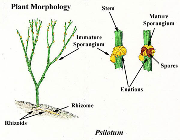
| Psilotum sporophyte anatomy |
As is the pattern in vascular plants, the diploid sporophyte generation is the dominant phase of the life cycle. Psilotophytes have many features that are similar to primitive vascular plants.
Like fossil vascular plants, psilotophytes have a horizontal stem called a rhizome. Aerial branches grow from the rhizome. The lower portions of the aerial stems in Psilotumare often five-sided,while the upper aerial stems are usually three-sided.
Both the rhizome and the aerial stem branch dichotomously, which means they tend to produce two equal branches at each node. Psilotum usually produces several dichotomies, or branching series, while Tmesipteris may have only one dichotomy on a particular aerial stem. Dichotomous branching is considered a primitive characteristic.
The outermost layer of the stem is composed of epidermal tissue. A waxy cuticle covers the epidermis of the aerial branches. Stomata are present in spaces between the ribs, which run length wise along the branch.
Interior to the epidermis is a wide cortex region. The cortex is composed of parenchyma cells. The cortex parenchyma is important for storage, as is evident from the presence of the many starch granules in each cell. Lying inward from the cortex in Psilotum is a layer of cells called the endodermis.
   |
Endodermal cells have a layer of suberin (fatty material) called a Casparian strip embedded in part of their cell walls to restrict water movement into and out of the vascular cylinder. The endodermis is well developed throughout the aerial part of the Psilotum plant body as well as the underground parts.
In Tmesipteris, the endodermis is present only in the rhizome. Alayer of cells containing tannins and phenolic compounds lies between the phloem cylinder and the cortex in Tmesipteris stems. This layer may be the physiologically active equivalent of an endodermis.
Although psilotophytes lack true roots, they do possess root like epidermal extensions called rhizoids. The rhizoids aid in absorption of water and mineral nutrients and also act as the points of entry for symbiotic fungi, whose presence may be essential for the survival of the organism.
The association of mutualistic fungi with plant roots or rhizoids is called a mycorrhiza. Because the fungi invade the body of the plant, they are sometimes referred to as endophytic fungi.
The xylem tissue of psilotophytes is composed of tracheids. Phloem tissue is made up of sieve cells, along with some parenchyma helper cells called albuminous cells. The stele, or vascular cylinder, of the psilotophyte rhizome is usually interpreted as a type of protostele.
Protosteles have no pith; they have a solid core of vascular tissue. In the protostelic actinostele of Psilotum, the solid core of xylem has arm-like extensions that reach into the surrounding tissues. Phloemsurrounds the xylem. In the lower portions of the aerial branches of Psilotum, the stele is considered to be a type of siphonostele.
In this case, the center of the stele is interpreted as a pith made up of sclerotic (hard, thick-walled) cells. This is similar to the aerial stem pattern in Tmesipteris. The tips of the aerial branches in Psilotum are strictly actinosteles.
Psilotum lacks true leaves. The leaflike appendages of the Psilotum shoot are called enations, or prophylls. The prophylls are composed of small flaps of photosynthetic tissue.
Small traces of vascular tissue end at the base of the prophyll and do not actually enter the structure. In contrast, the foliar appendages of Tmesipteris are larger and are considered to be a type of true leaf called a microphyll. Microphylls have a single vein extending into the blade of the leaf.
Spores are produced in sporangia located on very short shoots on the sides of aerial branches. The lateral placement of sporangia in psilotophytes contrasts with the terminal placement of sporangia in primitive vascular plants such as Rhynia.
Because each sporangium appears to represent a fusion of two sporangia (as in Tmesipteris) or three sporangia (as in Psilotum), they are generally called synangia.
Cells within the synangium undergo meiosis to produce haploid spores. The spores are described as monolete, meaning that they have a single ridge. The monolete character is considered a derived trait, which is different from the trilete, or three-ridged, spores of primitive vascular plants.
Gametophyte Anatomy
The haploid psilotophyte gametophytes are very small and grow underground. In general, they are cylindrical, brown in color, covered with rhizoids, and are often branched. The branching may be dichotomous, as in the sporophyte, or irregular in response to the wounding of the gametophyte’s apical meristems.
Gametophytes rely on endophytic fungi to obtain energy. Fungi invade nearly all of the cells of the gametophyte body except the apicalmeristems and the gametangia, or gamete-producing organs.
Both male and female sex organs are found scattered throughout a single plant. The antheridia (male organs) are small, multicellular, hemispherical structures that protrude from the surface of the gametophyte.
Cells inside the antheridia undergo mitosis to produce multiflagellated sperm cells. The archegonia (female organ) has a swollen base called the venter. The venter is sunken below the surface of the gametophyte.
A cell within the venter undergoes mitotic cell division to produce an egg. Four rows of cells form the neck of the archegonium, which surrounds a neck canal. Two cells initially fill the neck canal. These neck canal cells break down when the archegonium is ready for fertilization.