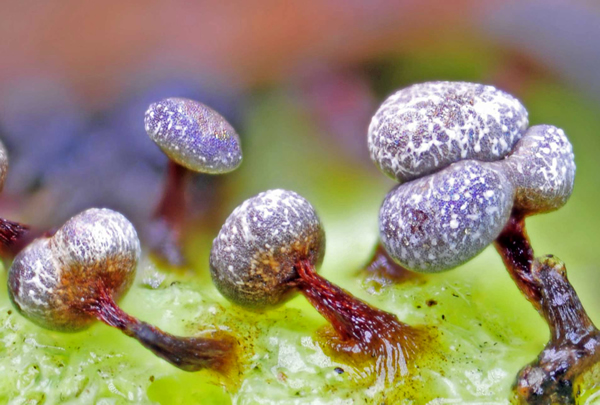
 |
| Plasmodial Slime Molds |
The plasmodial slimemolds, or myxomycetes, phylum Myxomycota, are a group of fungus-like organisms usually present and sometimes abundant in terrestrial ecosystems. However, this group comprises about eight hundred species, related neither to cellular slime molds nor to fungi.
The plasmodial slime molds have no cell walls and exist as thin masses of protoplasm, which appear to be streaming in a fan like shape, under favorable conditions.
As these masses, called plasmodia, travel, they absorb small particles of decaying plant and animal matter as well as bacteria, fungi, and yeasts. When mature, a plasmodium may weigh 20-30 grams and take up an area of 1 meter or more.
  |
Myxomycetes have been known fromtheir fruiting bodies (often a sporangium sprouting from a small mound that forms when the plasmodium stops moving) since at least the middle of the seventeenth century. Their life cycle has been understood for more than a century.
The reproductive, spore producing stage in the life cycle can achieve macroscopic dimensions and be collected and preserved for study in much the same way as mushrooms and other fungi or even specimens of bryophytes, lichens, and vascular plants.
However, most species of myxomycetes tend to be rather inconspicuous or sporadic in their occurrence and thus not always easy to detect in the field. Moreover, fruiting bodies ofmost species are relatively ephemeral and do not persist in nature for very long.
Myxomycetes also spend a portion of their life cycle as true eukaryotic microorganisms, when their very presence in a given habitat can be exceedingly difficult, if not impossible, to determine.
Life Cycle
 |
| Myxomycetes life cycle |
The life cycle of a myxomycete involves two very different trophic (or feeding) stages, one consisting of uninucleate (single-nucleus) amoeboid cells, with or without flagella, and the other consisting of a distinctive multinucleate structure, the plasmodium.
Under favorable conditions, the plasmodium gives rise to one or more fruiting bodies containing spores. The fruiting bodies produced by myxomycetes are somewhat suggestive of those produced by some fungi, although they are considerably smaller (usually no more than 1-2 millimeters tall).
The spores of myxomycetes are for most species apparently wind-dispersed and complete the life cycle by germinating to produce the uninucleate amoeboid cells. These cells feed and divide by binary fission to build up large populations in the various habitats in which these organisms occur.
The transformation from one trophic stage to the other in the myxomycete life cycle is in most cases the result of fusion between compatible amoeboid cells,which thus function as gametes.
   |
The fusion of the two cells produces a diploid zygote that feeds, grows, and undergoes repeated mitotic nuclear divisions to develop into the plasmodium. Bacteria represent the primary food resource for both trophic stages, but plasmodia are also known to feedupon yeasts, cyanobacteria, and fungal spores.
Myxomycete plasmodia usually occur in situations in which they are relatively inconspicuous, but careful examination of the inner surface of dead bark on a fallen log or the lower surface of a piece of coarse woody debris on the ground in a forest, especially after a period of rainy weather, often will turn up an example or two.
Most of the plasmodia encountered in nature are relatively small, but some species are capable of producing a plasmodium that can reach a size of more than 1 meter across.
Under adverse conditions, such as drying out of the immediate environment or low temperatures, a plasmodium may convert into a hardened, resistant structure called a sclerotium, which is capable of reforming the plasmodium upon the return of favorable conditions.
Moreover, the amoeboid cells can undergo a reversible transformation to dormant structures called microcysts. Both sclerotia and microcysts can remain viable for long periods of time and are probably very important in the continued survival of myxomycetes in some habitats, such as deserts.
Structure of Fruiting Bodies
 |
| myxomycetes fruiting bodies |
Identification of myxomycetes is based almost entirely upon features of the fruiting bodies produced by these organisms. Fruiting bodies (also sometimes referred to as “sporophores” or “sporocarps”) occur in four generally distinguishable forms or types, although there are a number of species that regularly produce what appears to be a combination of two types.
The most common type of fruiting body is the sporangium, which may be sessile or stalked,withwide variations in color and shape.
The actual spore-containing part of the sporangium (as opposed to the entire structure, which also includes a stalk in those forms characterized by this feature) is referred to as a sporotheca. Sporangia usually occur in groups, because they are derived from separate portions of the same plasmodium.
Asecond type of fruiting body, an aethalium, isa cushion-shaped, sessile structure. Aethalia are presumed to be masses of completely fused sporangia and are relatively large, sometimes exceeding several centimeters in extent.
A third type is the pseudoaethalium (literally, a false aethalium). This type of fruiting body, which is comparatively uncommon, is composed of sporangia closely crowded together.
Pseudoaethalia are usually sessile, although a fewexamples are stalked. The fourth type of fruiting body is called a plasmodiocarp. Almost always sessile, plasmodiocarps take the form of the main veins of the plasmodium from which they were derived.
A typical fruiting body consists of as many as six major parts: hypothallus, stalk, columella, peridium, capillitium, and spores.Not all of these parts are present in all types of fruiting bodies.
The hypothallus is a remnant of the plasmodium sometimes found at the base of a fruiting body. The stalk (also called a stipe) is the structure that lifts the sporotheca above the substrate. As already noted, some fruiting bodies are sessile and thus lack a stalk.
The peridium is a covering over the outside of the sporotheca that encloses the actual mass of spores. It may or may not be evident in a mature fruiting body. The peridium may split open along clearly discernible lines of dehiscence, as a preformed lid, or in an irregular pattern.
In an aethalium, the relatively thick covering over the sporemass is referred to as a cortex rather than a peridium. The columella is an extension of the stalk into the sporotheca, although it may not resemble the stalk.
The capillitium consists of threadlike elements within the sporemass of a fruiting body. Many species of myxomycetes have a capillitium, either as a single connected network or asmany free elements called elaters.
The elements of the capillitium may be smooth, sculptured, or spiny, or they may appear to consist of several interwoven strands. Some elements may be elastic, allowing for expansion when the peridiumopens,while other types are hygroscopic and capable of dispersing spores by a twisting motion.
Spores of myxomycetes are quite small and range in size from slightly less than 5 to occasionally more than 15 micrometers. Nearly all of them appear to be round, and most are ornamented to some degree. Spore size and also color are very important in identification. Spores can be dark or light to brightly colored.
Occurrence in Nature
There are approximately eight hundred recognized species ofmyxomycetes, and these have been placed in six different taxonomic orders: Ceratiomyxales, Echinosteliales, Liceales, Physarales, Stemonitales, and Trichiales.
However, members of the Ceratiomyxales are distinctly different from members of the other orders, and many modern biologists have removed these organisms from the myxomycetes and reassigned them to another group of slime molds, the protostelids.
The majority of species of myxomycetes are probably cosmopolitan, and at least some species apparently occur in any terrestrial ecosystem with plants (and thus plant detritus) present. However, a few species do appear to be confined to the tropics or subtropics, and others have been collected only in temperate regions.
Compared to most other organisms, myxomycetes show very little evidence of endemism, with the same species likely to be encountered in any habitat on earth where the environmental conditions suitable for its growth and development apparently exist.
Although the ability of a plasmodium to migrate some distance from the substrate upon or within which it developed has the potential of obscuring myxomycete-substrate relationships, fruiting bodies of particular species of myxomycetes tend to be rather consistently associated with certain types of substrates.
For example, some species almost always occur on decaying wood or bark, whereas others are more often found on dead leaves and other plant debris and only rarely occur onwood or bark.
In addition to these substrates, myxomycetes also are known to occur on the bark surface of living trees, on the dung of herbivorous animals, in soil, and on aerial portions of dead but still-standing herbaceous plants.
The myxomycetes associated with decaying wood are the best known, because the species typically occurring on this substrate tend to be among those characteristically producing fruiting bodies of sufficient size to be detected in the field.
Many of the more common and widely known myxomycete taxa, including various species of Arcyria, Lycogala, Stemonitis, and Trichia, are predominantly associated with decaying wood.