 |
| Angiosperm Cells and Tissues |
Some cell types and tissues which are not found in any other groups of plants occur in angiosperms (flowering plants).
Angiosperms are a group of plants with seeds that develop within an ovary and reproductive organs in flowers. They are commonly referred to as flowering plants and represent the most successful group of plants on earth, with approximately 235,000 species.
Various cell types and tissues, many of which are not found in any other groups of plants, occur in angiosperms. These cells and tissues perform varied functions, which are very efficient compared to their counterparts in other plants. These include dermal, vascular (xylem and phloem), and ground tissues (such as parenchyma, collenchyma, and sclerenchyma).
  |
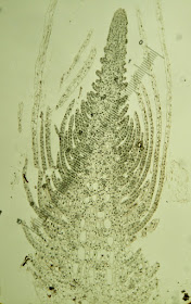
The growth of plants is carried on by a group of cells at their tips. These groups of cells are referred to as apical meristems, which are composed of initials and their most recent derivatives. The initials are the main source of body cells in plants,while the derivatives become any of the cells and tissues in the plant body.
The apical meristems of both the shoot and the root show continued cell division, with cells enlarging, elongating, and differentiating in regular, distinctly organized patterns. Apical meristems bring about the increase in the length of the stems and roots and are responsible in the formation of the primary plant body.
The shoot apical meristem may continually initiate the aerial components of the plant or may enter a state of periodic quiescence. In some plants, the shoot apical meristem transforms into a floral or inflorescence meristem that eventually terminates in a single flower or clusters of flowers, respectively.
The root apical meristem is enclosed by a thimble-shaped root cap that hastens the penetration of roots between soil particles.Unlike the shoot apical meristem, the root apical meristem forms no appendages. In fact, the site of lateral root initiation is far removed from it.
Shoot Apex
The shoot apical meristem is typically dome-shaped but flattened, and concave outlines also exist. The outline is not constant but changes in response to plastochron (the time interval between the initiation of one leaf primordium and the next). At least three models describe the shoot apical meristems. Although each of these is based on one or two unique criteria, they also have a few overlapping features.
Cell Lineage Analysis
This model holds that three clonally related layers of cells characterize the shoot apical meristem. These layers can be more than one cell layer thick. L1 is the outermost layer and gives rise to the epidermis, L2 is the middle layer and gives rise to the vascular tissues and cortex, and L3 is the inner most layer and gives rise to the pith.
This model was based on studies using periclinal chimeras (organs or parts of tissues of diverse genetic constitution), where one of the cell layers was genetically altered using drugs that inhibit separation of chromosomes.
Tunica-Corpus Concept
 |
| Tunica-Corpus Concept |
Partly enclosed by the tunica is the corpus, a group of loosely arranged cells that divide in various planes and give rise to the vascular and ground tissues. The tunica maintains its individuality by surface growth, whereas the corpus adds bulk by increase in volume.
Cytohistological Zonation
This model recognizes various definable zones in the shoot apical meristem. Three zone boundaries are distinguished by cell size: staining quality, degree of vacuolation, and frequency of cell division.
The central (mother cell) zone represents a conspicuous group of enlarged and isodiametric cells that undergo infrequent cell division, possess prominent nuclei, and are often highly vacuolated. The flanking peripheral zone is derived from, and partly surrounds, the central zone. Cells of this zone are smaller, are mitotically active, and have dense cytoplasms.
They give rise to the epidermis, vascular tissues, and cortex. The rib zone is located at the base of the central and peripheral zones. This zone is directly formed from the central zone, produces longitudinal files of cells by periclinal divisions, and gives rise to the pith.
Root Apex
 |
| Root Apex |
This organization shows a clear boundary between root cap and other tissues of the root apex. In an open type of organization, all of the root tissues share a group of initials, and therefore the boundary of the root cap is indistinguishable from the other tissues of the root apex.
Developmental Processes
The cells produced by apical meristems undergo several key developmental processes, which include growth, differentiation, and morphogenesis. Although each of these can be separated individually, they overlap in highly complex fashion.
Growth refers to the quantitative increase in a cell’s volume or mass due to enlargement and multiplication. Differentiation is the qualitative change in the form and function of organelles, cells, tissues, and organs, resulting in the establishment of new structures and functions.
From an anatomical point of view, cell differentiation is related to changes in cell size and shape, modifications of the wall, and changes in staining characteristics of nucleus or cytoplasm, as well as the degree of vacuolation and the ultimate loss of the protoplast in some cases. Morphogenesis is the visible manifestation of all of the changes, brought about by growth and differentiation, as expressed in the overall morphology of the plant.
Dermal Tissues
The primary plant body is composed of three basic tissues: dermal, vascular, and ground tissues. The dermal tissue (or epidermis) is made up of several cell types and is involved in a variety of functions, including retention and absorption of water and minerals, protection against herbivores, and control of gas exchange. Each of these functions is attributable to one or more of the unique features of the epidermis.
Most epidermal cells are flat and tightly packed, forming a single layer around stems, leaves, and other organs. The outer walls of epidermal cells are equipped with a waterproof layer made up of a fatty material called cutin. The tightly packed and cutinized epidermis protects the plants from desiccation by keeping moisture in.
Epidermal cells lack chloroplasts and are transparent. It is the underlying cells that give leaves and stems their green color. However, the vacuoles of some epidermal cells occasionally contain pigments and are responsible in the coloration of flowers and colored parts of variegated leaves.
Stomata are specialized structures that form part of the epidermis of leaves, stems, flowers, and fruits. They are involved in regulating the intake of carbon dioxide for photosynthesis as well as the release of oxygen. Trichomes are single-celled or multicellular out growths of epidermal cells that are involved in deterring herbivores and restricting transpiration.
Root hairs are also outgrowths of epidermal cells that are specialized for absorbing water and minerals from soil. They occur near the tip of the root and function to increase its absorptive surface area several-thousand fold.
Vascular Tissues
 |
| Vascular Tissues |
Vascular tissues are of two types: xylem and phloem. Xylem occurs throughout the plant body, and the type that differentiates directly from the apical meristem is called primary xylem. (Secondary xylem is formed from the vascular cambium.)
Primary xylem is formed as stems and roots elongate. The two kinds of conducting cells in xylem are tracheids and vessels, or vessel elements. Both are dead at maturity and have thick, lignified secondary cell walls. Tracheids are long, slender cells with tapered, overlapping ends.
They are the only water-conducting cells in most gymnosperms (an evolutionary line of plants that includes conifers).Water moves upward in roots and stems from tracheid to tracheid through thin areas in their cellwalls called pits. With only a few exceptions, all angiosperms contain vessel elements and tracheids.
Vessel elements are short, wide in diameter, and connected end to end. Their end walls are partly or wholly dissolved, forming long hollow vessels through which water moves. All these features of vessels enable them to transport water more rapidly than tracheids.
Phloem transports dissolved organic materials throughout the plant. The conducting cells of the phloem are called sieve elements, which are devoid of nuclei but otherwise have intact cytoplasm. They also have thin areas along their cell walls called sieve areas that are perforated. Solutes move from sieve element to sieve element through these pores.
Ground Tissues
The three types of ground tissue are parenchyma, collenchyma, and sclerenchyma. Parenchyma cells are the most abundant and versatile cells in plants. These cells are isodiametric, are alive at maturity, are highly vacuolated, and have a primary cell wall. Parenchyma functions as food-and-water-storage tissue as well as sites of metabolism in plants. Chlorenchyma cells are chloroplast-containing parenchyma specialized for photosynthesis.
Collenchyma cells are relatively long, with unevenly thickened primary walls. They support growing regions of shoots and are common in petioles, elongating stems and expanding leaves. Collenchyma cells are well adapted for support because their cell walls are able to stretch. They often form in strands or a cylinder just beneath the epidermis; such location maximizes support, as would a rod located in the center of a stemor petiole (leaf base).
Sclerenchyma cells are rigid; produce thick, non-stretchable secondary walls; and are usually dead atmaturity. They occur in, support, and strengthen mature regions of plants, including stems, roots, and leaves. There are two types of sclerenchyma cells: sclereids and fibers.
Sclereids are relatively short and variable in shape and usually occur in small groups. Fibers are long and slender and occur in strands or bundles. Sclereids are found in the roots, leaves, and stems.
They produce the gritty texture of pears and mostly make up the tough core of apples as well as the seed coats of peanuts and walnuts. Fibers are often associated with vascular tissues and, compared to sclereids, are typically elongated cells that vary in length from a few millimeters to more than half a meter long.

